Anatomy Of Chest : Frankentsein Diary With Human Anatomy Chest Vector Image. The anatomic illustrations are presented as… Anatomy of the thorax, heart, abdomen and pelvis recommended text gray's anatomy for students. Computed tomography (ct) of the chest can detect pathology that may not show up on a conventional chest radiograph(1). Muscles of the chest and their functions you have two mighty muscles on both sides of your chest: Chest a man's chest — like the rest of his body — is covered with skin that has two layers.
The chest or thorax is the region between the neck and diaphragm that encloses organs, such as the heart, lungs, esophagus, trachea, and thoracic diaphragm. Chest workoutschest workout routinechest workouts for masschest workouts at homechest workout bodybuildingchest workouts for men at homechest workout with du. First i'll do an intro to the different organs and structures in the chest, and then i'll go over some images showing their locations. The myotomes elongate and invade the mesoderm of the wall of the embryonic thoracic and abdominal cavities. The chest wall is comprised of skin, fat, muscles, and the thoracic skeleton.
1 from Thank you for visit anatomynote.com. Chest a man's chest — like the rest of his body — is covered with skin that has two layers. Anatomy of the thorax, heart, abdomen and pelvis recommended text gray's anatomy for students. Computed tomography (ct) of the chest can detect pathology that may not show up on a conventional chest radiograph(1). However, the classical anatomical descriptions in textbooks make it difficult to gain full mastery of this subject, because the books usually deal with its elements separately. It is enclosed by the ribs, the vertebral column, and the sternum, or breastbone, and is separated from the abdominal cavity (the body's largest hollow space) by a muscular and membranous partition, the diaphragm. The chest is made up primarily of two muscles: This is an eps 10 vector illustration and includes a high resolution jpeg.
Anatomically, the heart is located in the anterior thoracic cavity;
12 cm (5 in) in length, 8 cm (3.5 in) wide, and 6 cm (2.5 in) in thickness. Basic thoracic anatomy and physiology an understanding of thoracic imaging requires knowledge of the anatomy being imaged, as described in this chapter, as well as the imaging techniques applied to the thorax, covered in chapter 2. The circulatory system does most of its work. The chest or thorax is the region between the neck and diaphragm that encloses organs, such as the heart, lungs, esophagus, trachea, and thoracic diaphragm. Learn about each of these muscles, their locations, functional anatomy and exercises for them. In insects, crustaceans, and the extinct trilobites, the thorax is one of the three main divisions of the creature's body, each of which is in turn composed of multiple segments. The myotomes elongate and invade the mesoderm of the wall of the embryonic thoracic and abdominal cavities. These myotomes divide into the epimere and the hypomere. About the 6th week, the somites differentiate into the sclerotomes and the dermatomyotomes. The chest or thorax region of the upper body has a number of important organs that reside within it that may present with chest pain if they become compromised in. The muscles of the chest develop from the somites found in the mesoderm. Anatomically, the heart is located in the anterior thoracic cavity; Related posts of anatomy of the chest and stomach anatomy of the eye.
It provides protection to vital organs (eg, heart and major vessels, lungs, liver) and provides stability for movement. A line is drawn from anterior surface of the body of 6th thoracic vertebrae passing through the apex of the heart up to anterior lower most part of diaphragm. Chest workoutschest workout routinechest workouts for masschest workouts at homechest workout bodybuildingchest workouts for men at homechest workout with du. (1) the pectoralis major, and (2) the pectoralis minor. The chest or thorax is the region between the neck and diaphragm that encloses organs, such as the heart, lungs, esophagus, trachea, and thoracic diaphragm.
Figure 7 From Relevant Surgical Anatomy Of The Chest Wall Semantic Scholar from d3i71xaburhd42.cloudfront.net The chest wall is comprised of skin, fat, muscles, and the thoracic skeleton. A good radiologist knows the anatomy because knowing where structures normally live and recognizing the location of an abnormality helps to make or narrow the differential diagnosis. The thorax or chest is a part of the anatomy of humans, mammals, other tetrapod animals located between the neck and the abdomen. Muscles of the chest and their functions you have two mighty muscles on both sides of your chest: How to view the anatomical labels. It provides access to ct images in the axial plane, allowing the user to learn and review the lung anatomy interactively. Anatomynote.com found chest bone, ribs, lung, heart, xiphoid process, sternum anatomy from plenty of anatomical pictures on the internet. Summary:for adequate treatment of patients with breast cancer, mastologists should have a complete understanding of the anatomy of the thoracic wall, axilla and breast.
The chest is made up primarily of two muscles:
Anatomy of the thorax, heart, abdomen and pelvis recommended text gray's anatomy for students. We think this is the most useful anatomy picture that you need. It provides access to ct images in the axial plane, allowing the user to learn and review the lung anatomy interactively. A good radiologist knows the anatomy because knowing where structures normally live and recognizing the location of an abnormality helps to make or narrow the differential diagnosis. Related posts of anatomy of the chest area anatomy of rib cage. Browse 2,533 female chest anatomy stock photos and images available, or start a new search to explore more stock photos and images. (1) the pectoralis major, and (2) the pectoralis minor. It provides protection to vital organs (eg, heart and major vessels, lungs, liver) and provides stability for movement. A typical heart is approximately the size of your fist: Swensen fund for innovation in teaching. Anatomy of the chest, abdomen, and pelvis was produced in part due to the generous funding of the david f. The epidermis is the outermost layer that provides a protective, waterproof seal over the body. The chest anatomy includes the pectoralis major, pectoralis minor and the serratus anterior.
The anatomic illustrations are presented as… Anatomy of the chest, abdomen, and pelvis was produced in part due to the generous funding of the david f. The chest is made up primarily of two muscles: The chest or thorax is the region between the neck and diaphragm that encloses organs, such as the heart, lungs, esophagus, trachea, and thoracic diaphragm. The circulatory system does most of its work.
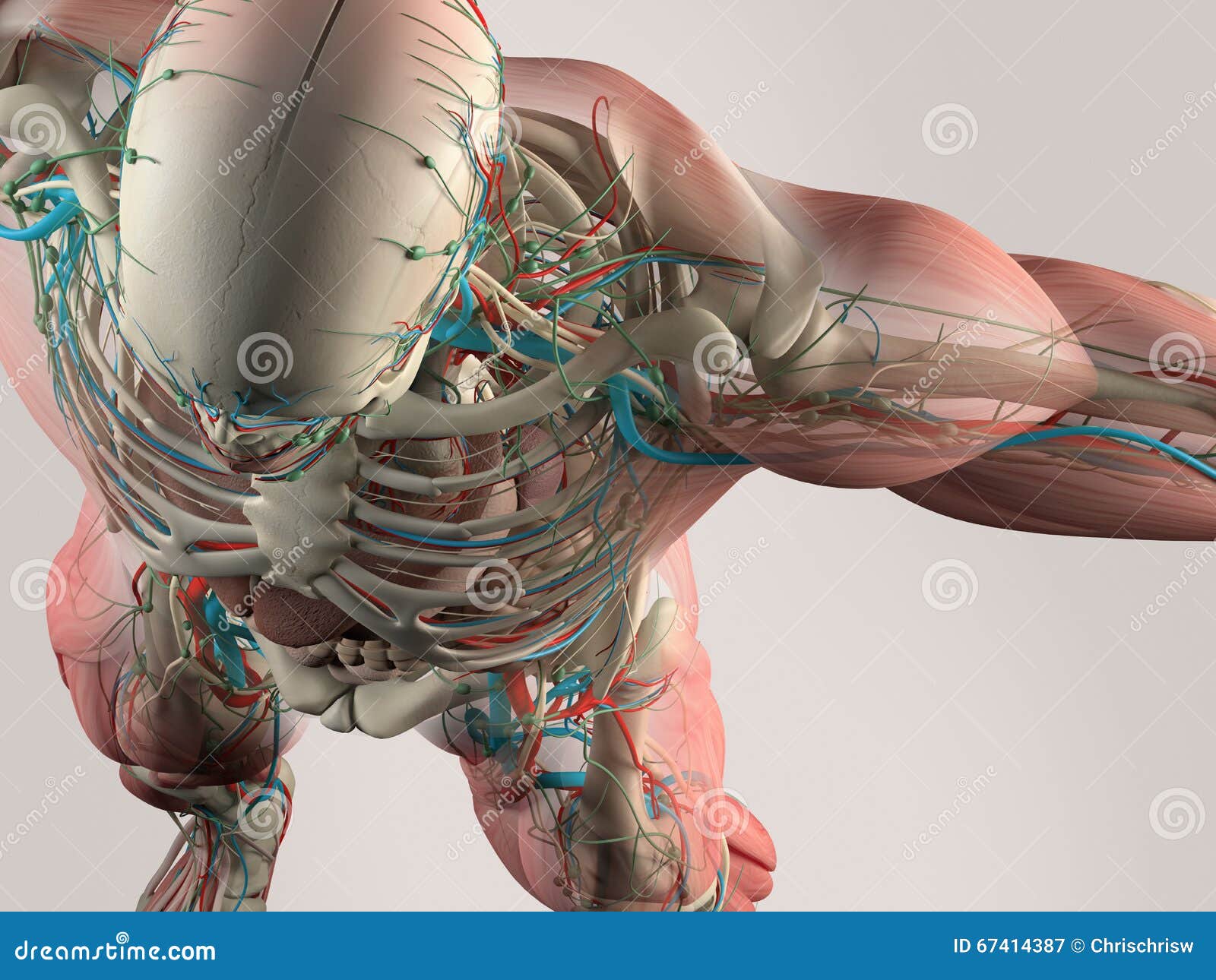
Human Anatomy Detail Of Chest And Shoulder Muscle Arteries On Plain Studio Background Human Anatomy Detail Of Skull And Shoulde Stock Illustration Illustration Of Male Muscles 67414387 from thumbs.dreamstime.com Basic thoracic anatomy and physiology an understanding of thoracic imaging requires knowledge of the anatomy being imaged, as described in this chapter, as well as the imaging techniques applied to the thorax, covered in chapter 2. The chest wall is comprised of skin, fat, muscles, and the thoracic skeleton. The circulatory system does most of its work. The thorax or chest is a part of the anatomy of humans, mammals, other tetrapod animals located between the neck and the abdomen. Thoracic cavity, also called chest cavity, the second largest hollow space of the body. 12 cm (5 in) in length, 8 cm (3.5 in) wide, and 6 cm (2.5 in) in thickness. Summary:for adequate treatment of patients with breast cancer, mastologists should have a complete understanding of the anatomy of the thoracic wall, axilla and breast. It provides access to ct images in the axial plane, allowing the user to learn and review the lung anatomy interactively.
However, the classical anatomical descriptions in textbooks make it difficult to gain full mastery of this subject, because the books usually deal with its elements separately.
Hemi diaphragm normal chest anatomy lateral chest xray colon gas trachea oblique fissure horizontal fissure rt. These myotomes divide into the epimere and the hypomere. Swensen fund for innovation in teaching. The muscles of the chest develop from the somites found in the mesoderm. You can click the image to magnify if you cannot see clearly. (1) the pectoralis major, and (2) the pectoralis minor. Anatomy of the eye 12 photos of the anatomy of the eye anatomy of the eye bones, anatomy of the eye lacrimal gland, anatomy of the eye special senses vision, anatomy of the eye ultrasound, external anatomy of the eye quiz, human anatomy, anatomy of the eye bones, anatomy of the eye lacrimal gland, anatomy … Anatomy of the chest, abdomen, and pelvis was produced in part due to the generous funding of the david f. The pectoralis major and the pectoralis minor, known collectively as your pecs. Radiology basics of chest ct anatomy with annotated coronal images and scrollable axial images to help medical students and junior doctors learning anatomy. Related posts of anatomy of the chest and stomach anatomy of the eye. The chest anatomy includes the pectoralis major, pectoralis minor and the serratus anterior. Anatomy of the thorax, heart, abdomen and pelvis recommended text gray's anatomy for students.
Share :
Post a Comment
for "Anatomy Of Chest : Frankentsein Diary With Human Anatomy Chest Vector Image"


Post a Comment for "Anatomy Of Chest : Frankentsein Diary With Human Anatomy Chest Vector Image"